
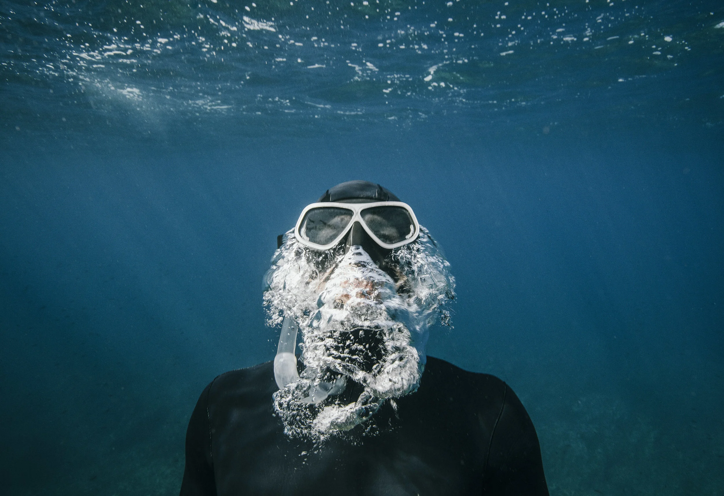
Diving Safety, Ear Barotrauma and Decompression Sickness
Having worked as both an Emergency Doctor and a Dive Doctor I know that diving, when not done correctly, can be very dangerous but when done safely it is the most wondrous thing; the sense of adventure and exploration can be overwhelming. It is important that people are trained and educated in all safety procedures and know how to look after themselves and each other. Here I will cover some of the major topics, including recognition, prevention and management. The sections include;
Decompression Sickness
Barotrauma, including damage to ears, sinuses and lungs
Outer Ear Infection
Medical Screening And Examination For Fitness To Dive
Resources
References
If you have any suggestions for additions or updates please leave a comment below.
MB ChB, College of Emergency Medicine Certificate Of Training In Good Clinical Practice In Research
Decompression Sickness
Decompression sickness (DCS) is also known as 'the bends' or caisson disease. It occurs when gas which has been dissolved in the blood comes out of solution and turns into bubbles inside the body. This happens usually during ascent when the pressure drops. It is similar to when the lid of a fizzy drink bottle is loosened and the pressure released; this is when the bubbles form. It can also happen during other periods of decompression such as flying in an aeroplane which is unpressurised at altitude.
The risk is reduced by following simple decompression techniques. This means that the condition does not occur often. When it does occur it can have life-changing consequences meaning that people take it very seriously. As a result most divers use a dive computer or table to measure their exposure and manage their decompression in order to avoid problems. It is recommended that's divers ascend at about 10 m per minute. Decompression stops may be scheduled, depending on the dive. Many divers will schedule a brief safety stop at 3m, 4.6 m or 6 m, depending on their training regime.
The schedules are taken from dive computers, software or tables. These use an algorithm to model gas exchange in the body.
Incidence
DCS is rare, with an estimated incidence of 2.8 per 10000 dives. This approximates to 1000 divers in the US every year.

A hyperbaric chamber.
Treatment
Patients suspected of DCS should be given 100-percent oxygen to breathe as soon as possible. A non-rebreather mask or other specialised gas delivery system should be used to maximise oxygen delivery. Mild cases may settle down spontaneously. Signs and symptoms involving the brain, heart and spinal cord should be managed with hyperbaric oxygen. A treatment which has been shown to be effective for minor DCS is recompression on air. Usually, recompression occurs in a recompression chamber using oxygen. A higher risk option is in-water recompression.
Oral or intravenous fluids should be given to treat dehydration and other life support measures should be taken. Recompression duration is dependent upon response to treatment as well as severity signs.
Pathophysiology
The formation of bubbles is thought to cause temporary blockage of arteries, capillaries and veins. The signs, symptoms and any subsequent damage will depend on the tissues supplied by the blocked vasculature. The lack of blood flow means that the tissues are starved of oxygen and tissue death and necrosis may occur. This can cause pain and failure of the tissue and organ.
Bubbles can trigger the formation of a blood clot with aggregation of platelets and activation of the clotting cascade. Damage to the lining of vessels (endothelium) may occur.
Signs and symptoms.
Joints
The commonest site for signs and symptoms are the elbows, ankles, knees and shoulders. Joint pain ('the bends') occurs in about two-thirds of occurrences. The shoulder is the most usual site. Pain can range from slight to unbearable, tends to be localised and characterized by a dullness, rather than a sharp pain. Pains tend to be deep.
Skin
When the skin is involved it becomes itchy, mottled and swollen. This tends to be in the upper parts of the body. Formication may occur, this is the feeling of ants walking on the skin.
Central nervous system
Headache and visual disturbances are signs of neurological involvement and this occurs in about 10% of patients. Other signs include altered vision, sensation, behaviour and mood. Memory loss, confusion and in serious cases, seizures and coma may also be part of the condition.
Spinal cord
If the spinal cord is involved there may be incontinence of urine and faeces. Paralysis or weakness in the legs and a tightening feeling around the abdomen and chest known as 'girdling' can also happen.
Lung
Lung involvement is very rare. When it occurs it results in a dry cough, shortness of breath and chest pain. The pain is pleuritic in nature, meaning that it is worse on coughing or deep inspiration.
Inner ear
Involvement of the inner ear affects the hearing and balance system. It results in loss of balance, dizziness, vomiting, vertigo (a feeling of spinning) and deafness.
Onset of symptoms
Although symptoms may occur immediately after diving in the majority of cases symptoms do not appear for at least an hour. It is important for Dive Physicians to be aware that symptoms may not occur for up to 48 hours. This can make diagnosis tricky; a diver with a painful elbow may have sprained it lifting her tank but has she got the bends? More on this later.
Causes
Decompression sickness is caused by a pressure drop, resulting in bubbles forming in the tissues. This is usually caused by leaving an area of high pressure and entering a lower pressure area. It is best known for affecting divers during their ascent. It occurs due to the fact that divers breathe gas which is at a higher pressure than atmospheric pressure. This causes the gas to dissolve in the blood at high pressure meaning it can escape suddenly as bubbles, if the pressure drops too quickly.
It is the inert gases that are inhaled that caused the problem. The majority of standard air consists of nitrogen but when diving helium can also cause DCS if it is in the tank.
Risk factors
DCS is more likely in longer dives at deeper depths which is logical but most risk factors are not fully understood. It appears that some people may be more susceptible than others but it is not understood why. It can be prevented by slow, staged decompression, allowing the gas to be released safely. DCS does not occur on descending a mountain as the pressure change is to slow. It was commonplace after high altitude plane flights in the 1930s until the advent of pressurized cabins. Despite this, divers who ascend a mountain in a car or take a flight are at higher risk. Surprisingly, cabin pressure on commercial airlines may only be 73% of atmospheric pressure. This increases the risk of the bends in people who have recently been diving.
A significant study showed no link between risk of DCS when diving and asthma, diabetes, heart disease, smoking or obesity. A higher risk was found with increased depth and previous decompression illness. A higher consecutive number of days of diving and being a man were also associated with increased risk. A lower risk was found for people using drysuits and nitrox. Higher diving frequency over the previous year, older age and more years since qualification also helped.
A greater rate of ascent, dehydration and previous injury increase the risk, as does getting very cold. Although alcohol can cause dehydration, a study in 2005 demonstrated no link.

Diagram of an atrial septal defect, this causes the blood to flow the wrong way in patent foramen ovale.
Patent Foramen Ovale
Interestingly, this heart defect can put people at higher risk of serious injury from DCS. A defect between the upper atrial heart chambers allows blood to flow in the wrong direction if the diver coughs or has raised pressure in the chest. This can cause blood containing bubbles which would normally be filtered out by the lungs to reach the heart or cause an arterial gas embolism. Serious injury can occur. Usually the lungs would filter these harmlessly.
Diagnosis
A doctor caring for divers needs to have a high index of suspicion for decompression sickness. The diagnosis must be considered if any of the symptoms or signs are demonstrated within a few hours of decompression. The less time between decompression and the onset of signs and symptoms, the more likely the condition is the cause. Recompression resulting in resolution confirms the diagnosis. The bubbles often show on a CT or MRI scan but the history of events is better at determining the diagnosis.
Prognosis
If a patient is immediately given 100% oxygen before recompression they will usually suffer no permanent effects. Long-term life-changing injury is possible. A study in 1987 followed sufferers up after three months. This showed 21.3% of 268 divers had ongoing symptoms. 16% of patients had permanent brain or spinal cord damage on long-term follow-up.
Barotrauma
Barotrauma is damage to tissues and organs as a result of a pressure gradient. Damage occurs due to stretch and shear. This can result in mechanical failure of the tissue and release of gas and other tissue contents into the surrounding areas. This can result in pain and other signs and symptoms depending on the area affected.
Types Of Barotrauma Associated With Diving
Barotrauma commonly affects the middle ear, sinuses, lungs and other areas through DCS. It occurs in the same scenarios as decompression sickness, but can also occur with a pressure wave. These are associated with an explosion, other factors can also cause the problem.

Anatomy of the ear. The middle ear bones are in the middle ear cavity. If the Eustachian tube is blocked expansion of the air here may burst the tympanic membrane.
Ear Barotrauma
The inner, middle or external ear can be affected by barotrauma. The middle ear is the most commonly affected, probably because of the air space within it.
Tight fitting diving hoods and wax in the outer ear canal can cause barotrauma to the external ear. Pressure damage to the inner ear can cause deafness and vertigo. Hyperacusis (over-sensitivity to sound) can also occur.
People with DCS who are being treated in a hyperbaric chamber need to equalise the ear pressures to avoid ear barotrauma.
Sinus Barotrauma
The sinuses in the head are full of air and so are susceptible to pressure damage if the drainage channels become blocked. Bleeding from the nose and pain may occur.
Mask Squeeze
Scuba masks contain air with no ventilation route unless the mask is equalised. Otherwise bleeding can occur under the skin (petechiael hemorrhages). It can also occur in the surface of the eyes (subconjunctival hemorrhages).

Pneumothorax.
Pulmonary Barotrauma And Pneumothorax
Over-inflation injury of the lungs can occur on ascent if a diver hold his breath. This can be prevented by slowly breathing out, preventing an increase in the volume within the lungs. Ascending 10m causes a doubling of gas volume, easily enough to cause a pneumothorax without equalisation. Pneumothorax occurs when gas escapes from within the lung or bronchial tree (breathing tubes). It enters the potential space around the lung allowing it to collapse.
Pneumothorax can be life-threatening and may cause death almost immediately if not treated in time. Tension pneumothorax is more dangerous. It is more rapidly progressive and causes the heart and large vessels in the centre of the chest to shift laterally. This blocks blood flow and can lead rapidly to cardiac arrest in less than a minute. Divers who hold their breath and do not use gas tanks are not affected by pneumothorax. This is because they only have one lungful of air which re-expands to a volume similar to its original on ascent.
How To Prevent Barotrauma In Divers
Barotrauma in divers can be prevented by equalising the pressure across the tissues. The method used for this varies depending on the area of the body involved.
Equalization Of Pressure In The Ear And Sinuses
Without equalising the pressure is the middle ear a ruptured tympanic membrane, also known as burst eardrum, may occur. This is more common on ascent when the hydrostatic pressure acts to force the Eustachian tube closed, making it harder for air to escape. On descent the increasing pressure more easily causes air to escape down the Eustachian tube into the nose. This equalises the pressure. The risk of eardrum damage is increased in the case of upper respiratory tract infection (URTI), one type of which is commonly called a cold. Inflammation of the ear and Eustachian tube makes equalization more difficult. There are several methods to open the Eustachian tube and equalise the pressure. The commonest is to simply swallow. If this doesn't work holding the nose and pushing the jaw forwards at the same time can help.
Equalising The Pressure In The Lungs To Prevent Barotrauma
In order to equalise the pressure in the lungs on ascent all that needs to be done is to not hold the breath and to allow air to escape from the mouth.
Equalising The Pressure In A Diving Mask
This can be performed by simply blowing air through the nose into the mask. Eye goggles which do not cover the nose should not be used for deep diving because the air space inside them cannot be equalised.
Equalising Pressure In A Drysuit To Prevent Dry Suit Squeeze
Bruising and pinching are the main risk from the folds of a drysuit on descent. A low pressure supply of gas is often used to equalise the pressure gradient. Squeez is avoided by injecting air into the suit manually and then vented on ascent to control buoyancy.
How To Equalise Diving Helmet Pressure To Prevent Diving Helmet Squeeze
If the hose supplying gas to a diving helmet is cut and there is failure of the non-return valve on the helmet then diving helmet squeeze is possible. An accidental rapid descent may cause a temporary squeeze. These risks have been significantly reduced through the use of higher flow rates of gas and a non-return valve. If a neck dam is present it may let water in if the pressure inside drops too far. This causes only a minor problem in small amounts but clearly could be very dangerous if large amounts enter.
Treatments
Clearly the treatments of diving related barotrauma will vary depending on the location of the trauma and its severity. Pneumothorax may require a chest drain to let the air out of the chest cavity and allow the lung to re inflate. Needle decompression and thoracostomy are alternatives. Management of DCS and arterial embolism using high flow oxygen and hyperbaric chambers is discussed above.
As barotrauma can cause life-threatening injuries management needs to involve life support. This may include intubation, ventilation, chest compressions, fluids and other measures as necessary.
Squeeze of the middle ear and sinuses may be treated with anti-inflammatories and decongestants. Some divers will take these before diving if they have an upper respiratory tract infection in order to reduce the risk of ear barotrauma. This can help in some situations but may increase the risk of burst eardrum. The diver may manage to descend without problem and then be unable to equalise the pressure on ascent, resulting in barotrauma. Without the medications they may not have been able to equalise their ears on descent. This would prevent them descending to a level where significant damage could occur.
Pain may be managed with paracetamol and opiates such as codeine, Oramorph and intravenous morphine, if required.
Outcome
If a diver suffers barotrauma they should not dive until a dive doctor has cleared them. Significant ear damage may need to be assessed by an ENT surgeon. Most tympanic perforations will heal on their own but some may need a surgical repair. Recovery from pneumothorax may take weeks, patients should not fly until cleared by an experienced doctor.

Scuba divers are at risk of swimmers' ear.
Ear Infections
Otitis externa (swimmer's ear/outer ear infection) is common in those who spend time in the sea or pool, so scuba divers are at high risk. The longer people spend in the water the higher the chance of them developing otitis externa (outer ear infection). The water softens the skin and allows the bacteria it contains to penetrate into the underlying dermis. This causes inflammation and infection.
Soapy, warm and dirty water increase the risk further so should be avoided, especially once an infection has taken hold. Common signs and symptoms include, itchiness, pain, skin flaking, minor discharge and muffled hearing. This increases the risk of otitis media (middle ear infection) which puts the diver at high risk of perforated eardrum. Middle ear infection causes worse pain, hearing sounds like the head is underwater and fluid can be seen behind the drum using an otoscope.
Swimming ear plugs are an effective method of protecting against swimmers’ ear in swimmers but should never be used whilst diving. This is because they create an unventilated and therefore unequalisable air space between the eardrum and the plug. This can cause ruptured tympanic membrane.
Whilst working as a dive doctor in Honduras the sea water was very warm which meant that ear infections were common. An audit of the patients who consulted me on the island showed that over half of them had ear infections. Some of them were so desperate to dive they were still going in the water even when they had pus running from their ears and down their faces. Many of them managed the risk with high dose ibuprofen and decongestants. Unfortunately one lady suffered a ruptured eardrum; she cried when I told her she would be out of the water for the rest of the season.
Prevention
Prevention is better than cure! Rinsing the ears with drinking water and/or surgical alcohol after immersion reduces the risk. It is important to dry them and try to keep cool when out of the water in order to avoid Mediterranean Ear. This is otitis externa secondary to having sweaty ears in hot climates.
Treatment
Mild infections may settle down simply by keeping our of the water but once it becomes established antibiotics will be required. Drops containing an antibiotic such as gentamicin, a steroid such as prednisolone and an antifungal such as acetic acid (vinegar) may be prescribed. If the infection is severe then oral antibiotics may also be required.
Medical Screening And Examination For Fitness To Dive
Fitness to dive is assessed periodically in professional divers. They are screened for risk factors which, if found, may mean the diver is not allowed to dive. Recreational divers do not usually need a medical assessment. They do usually complete a statement to identify the most common and dangerous risk factors.
COPD (chronic obstructive pulmonary disease) increases the risk of pneumothorax. Asthma and Marfan syndrome also carry higher risk. These may or may not completely prevent a diver from diving, depending on the severity and local guidelines. In some situations asthmatics who are well controlled and have only mild disease may be able to dive. Relevant history and examination should be aimed at identifying the conditions mentioned in this article.
Resources
This article is not to be used as medical advice. You must consult your medical and diving professionals if any of the issues in this article are relevant to you.
Dr Toby Bateson
References
Image of hyperbaric chamber courtesy of Intermedichbo at sr.wikipedia [CC BY 3.0 rs (https://creativecommons.org/licenses/by/3.0/rs/deed.en)]
Image of atrial septal defect Manco Capac [CC BY-SA 3.0 (https://creativecommons.org/licenses/by-sa/3.0)]
Image of pneumothorax BruceBlaus. Blausen.com staff (2014). "Medical gallery of Blausen Medical 2014". WikiJournal of Medicine 1 (2). DOI:10.15347/wjm/2014.010. ISSN 2002-4436. [CC BY 3.0 (https://creativecommons.org/licenses/by/3.0)]
Image of anatomy of ear BruceBlaus [CC BY-SA 4.0 (https://creativecommons.org/licenses/by-sa/4.0)]
Pulley, Stephen A (27 November 2007). "Decompression Sickness". Medscape.
Francis, T James R; Smith, DJ (1991). "Describing Decompression Illness". 42nd Undersea and Hyperbaric Medical Society Workshop. 79(DECO)5–15–91.
U.S. Navy Supervisor of Diving (2008). "Chapter 20: Diagnosis and Treatment of Decompression Sickness and Arterial Gas Embolism". U.S. Navy Diving Manual(PDF). SS521-AG-PRO-010, revision 6. volume 5. U.S. Naval Sea Systems Command. p. 37. Archived from the original (PDF) on 5 March 2011.
U.S. Navy Supervisor of Diving (2008). U.S. Navy Diving Manual (PDF). SS521-AG-PRO-010, revision 6. vol.5. U.S. Naval Sea Systems Command. pp. 20–25. Archived from the original (PDF) on 5 March 2011.
Sheffield, Paul J; Vann, Richard D (2002). Flying After Diving Workshop. Proceedings of the DAN 2002 Workshop. United States: Divers Alert Network. p. 127. ISBN 978-0-9673066-4-3.
Moon, Richard E; Kisslo, Joseph (1998). "PFO and decompression illness: An update". South Pacific Underwater Medicine Society Journal. 28 (3). ISSN 0813-1988. OCLC 16986801.
Kindwall, Eric P; Baz, A; Lightfoot, EN; Lanphier, Edward H; Seireg, A (1975). "Nitrogen elimination in man during decompression". Undersea Biomedical Research. 2 (4): 285–297. ISSN 0093-5387. OCLC 2068005. PMID 1226586.
Divers Alert Network (1997). "Report on Diving Accidents and Fatalities in 1995". Divers Alert Network. Retrieved 23 May 2010.
US Navy Diving Manual, 6th revision. United States: US Naval Sea Systems Command. 2006.
Fitzpatrick, D. T.; Franck, B. A.; Mason, K. T.; Shannon, S. G. (1999). "Risk factors for symptomatic otic and sinus barotrauma in a multiplace hyperbaric chamber". Undersea and Hyperbaric Medicine. 26 (4): 243–7. PMID 10642071.
Butler, F. K.; Gurney, N. (2001). "Orbital hemorrhage following face-mask barotrauma". Undersea and Hyperbaric Medicine. 28 (1): 31–4. PMID 11732882.
Haake, Ronald; Schlichtig, Robert; Ulstad, David R.; Henschen, Ross R. (April 1987). "Barotrauma: Pathophysiology, Risk Factors, and Prevention" (PDF). Chest. 91(4): 608–613. doi:10.1378/chest.91.4.608
Lehm, Jan P.; Bennett, Michael H. (2003). "Predictors of middle ear barotrauma associated with hyperbaric oxygen therapy". South Pacific Underwater Medicine Society Journal. 33: 127–133.
Barsky, Steven; Neuman, Tom (2003). Investigating Recreational and Commercial Diving Accidents. Santa Barbara, California: Hammerhead Press. pp. 61, 90. ISBN 978-0-9674305-3-9.
Vorosmarti, J.; Linaweaver, P. G., eds. (1987). "Fitness to Dive. 34th Undersea and Hyperbaric Medical Society Workshop". UHMS Publication Number 70(WS-WD)5-1-87. Bethesda, Maryland: Undersea and Hyperbaric Medical Society.
Adir, Yochai; Bove, Alfred A. (2016). Yochai Adir and Alfred A. Bove (eds.). "Can asthmatic subjects dive?" (PDF). Number 1 in the Series "Sports-related Lung Disease". European Respiratory Review. 140 (140): 214–220. doi:10.1183/16000617.0006-2016. PMID 27246598.